
Imaging

Synaptic changes following learning are measured at the electron microscopic level using unbiased stereological approaches in collaboration with Yuri Geinisman and Dan Nicholson (Rush University Medical Center). From Geinisman et al. 2000
What does the development of memory formation look like and what is the nature of the interactions that exist between the hippocampus and cortical regions to enable information storage? How do the structures of cells change in response to learning? How does the influx of ions into cells differ as a consequence of learning? Imaging techniques that enable the visualization of functional and structural changes in the whole brain, individual cells, or select regions of cells are used to investigate these questions. Imaging procedures complement single neuron recording experiments and all other areas of research in the laboratory by allowing us the ability to visualize a memory from acquisition to consolidation. Human imaging experiments will explore the generalizability and translatability of our work.
Example of BOLD activation in response to right whisker stimulation in awake rabbits. Figure from Schroeder et al. Neuroimage, 2016.
Magnetic resonance imaging permits examination of the entire brain simultaneously and observation of changes in brain activity in the same individual over time. Functional magnetic resonance imaging is being done in rabbits with our collaborators Daniel Procissi and Alice Wyrwicz (7 and 4.7 Tesla magnet) and in humans together with the Northwestern Brain Mapping Group (3 Tesla magnet).
Representative publications:
- Schroeder MP, Weiss C, Procissi D, Wang L, Disterhoft JF (2016) Pretrial functional connectivity differentiates behavioral outcomes during trace eyeblink conditioning in the rabbit. Learn Mem. 23(4):161-8.
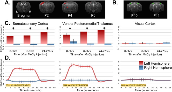
- Schroeder MP, Weiss C, Procissi D, Wang L, Disterhoft JF (2016) Activity-induced manganese-dependent MRI (AIM-MRI) and functional MRI in awake rabbits during somatosensory stimulation. Neuroimage 126:72-80.
- Geinisman Y, Disterhoft JF, Gundersen HJ, McEchron MD, Persina IS, Power JM, van der Zee EA, West MJ (2000). Remodeling of hippocampal synapses after hippocampus-dependent associative learning. J Comp Neurol 417(1): 49-59.
- Miller MJ, Weiss C, Song X, Iordanescu G, Disterhoft JF, Wyrwicz AM (2008). Functional magnetic resonance imaging of delay and trace eyeblink conditioning in the primary visual cortex of the rabbit. J Neurosci 28(19): 4974-4981.